Nuclear Lamin Molecular Weight
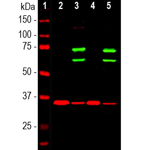
Red signal from loading control ab10799 observed at 18 kda.
Nuclear lamin molecular weight. Wild type hap1 cell lysate lane 2. Validated in wb ihc flow cyt and tested in mouse rat sheep hamster cow dog human. Lamin b1 knockout hap1 nuclear lysate 10 µg lanes 1 and 2. Use nuclear extracts incubate primary antibody overnight.
Lamin a and c are cleaved by caspases into large 41 50 kda and small 28 kda fragments which can be used as markers for apoptosis 4 5. Knockout tested mouse monoclonal lamin a antibody 133a2. The bands are representative of three independent experiments in triplicate for each protein and beta actin was used as a housekeeping gene. Ab16048 was shown to specifically react with lamin b1 in wild type hap1 cells.
66 kda lanes 1 4. Lmnb1 knockout hela cell lysate lysates proteins at 20 µg per lane. During apoptosis lamin a c is specifically cleaved into a large 41 50 kda and a small 28 kda fragment 3 4. Green signal from target ab16048 observed at 68 kda.
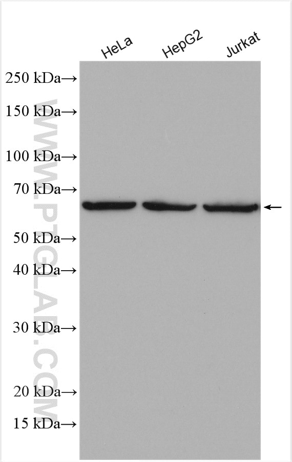
During mitosis the lamina matrix is reversibly disassembled as the lamin proteins are phosphorylated. 65 70 kda depending on the species. Lmnb1 knockout hap1 cell lysate lane 3. Merged signal red and green.
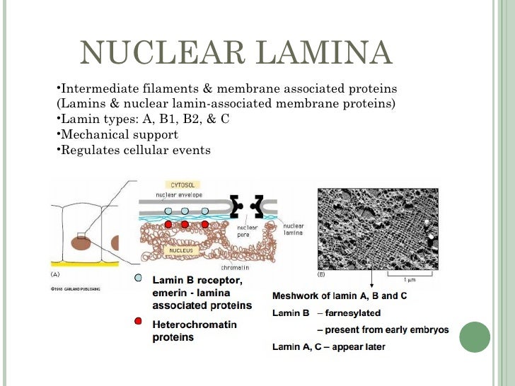
Lamin b1 is also cleaved by caspases during apoptosis 9. The lamins are type v intermediate filaments which can be categorized as either a type lamin a c or b type lamin b 1 b 2 according to homology of their dna sequences biochemical properties and cellular localization during the cell cycle. Cited in 63 publication s. The cleavage of lamins results in nuclear dysregulation and cell death 5 6.
Immunocytochemistry immunofluorescence anti lamin b1 antibody 119d5 f1 nuclear envelope marker ab8982 this image is courtesy of marilena ciciarello patrizia lavia university la sapienza. Green ab133741 observed at 70 kda. Vertebrate lamins consist of two types a and b. Anti lamin b1 antibody epr8985 b ab133741 at 1 1000 dilution lane 1.
Mouse monoclonal lamin a lamin c antibody 131c3 nuclear envelope marker. Type b lamins consist of lamin b1 and b2 encoded by separate genes 6 8. Wild type hap1 nuclear lysate 10 µg lane 2. Lamin a c is cleaved by caspase 6 and serves as a marker for caspase 6 activation.
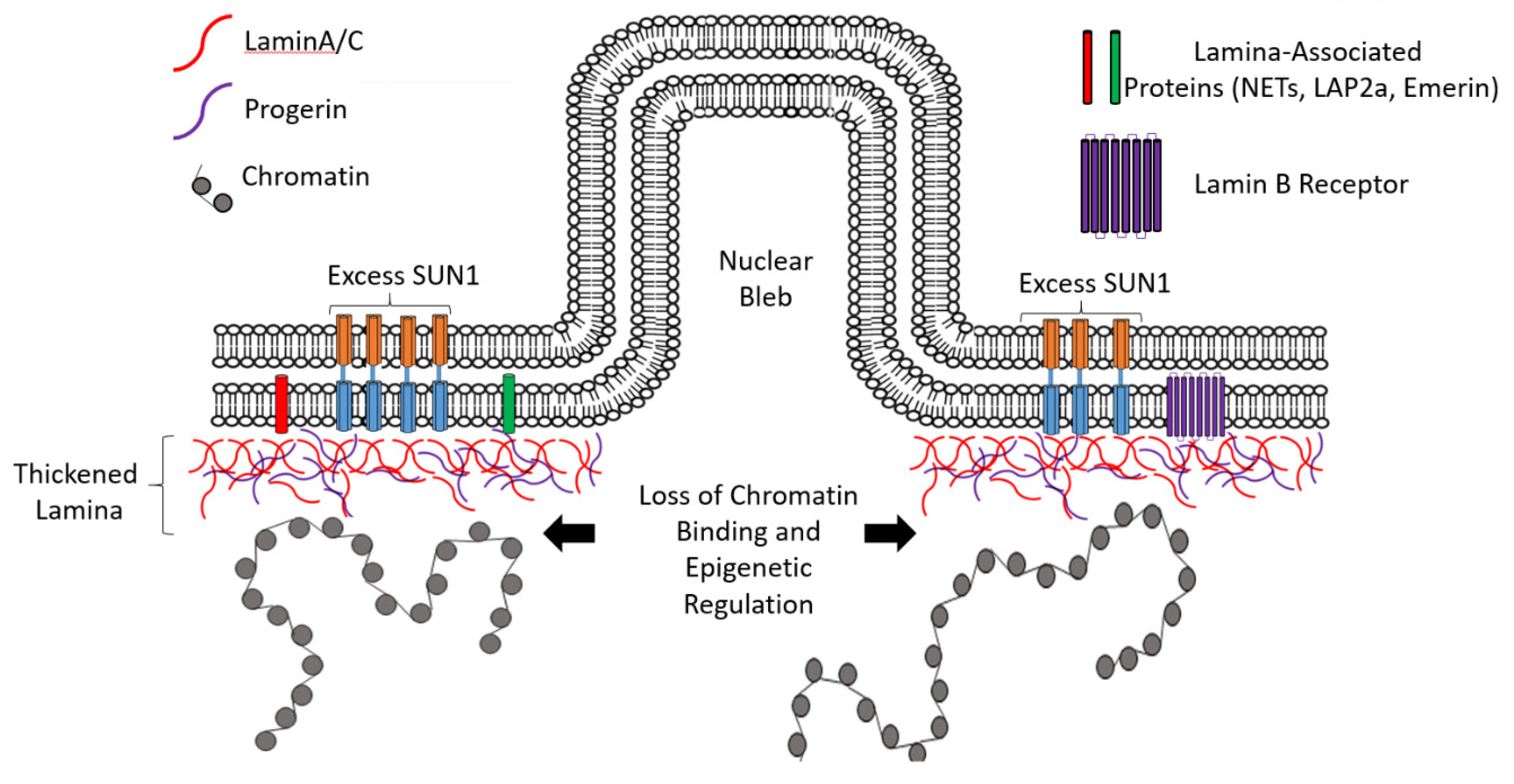
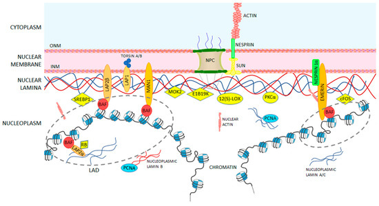
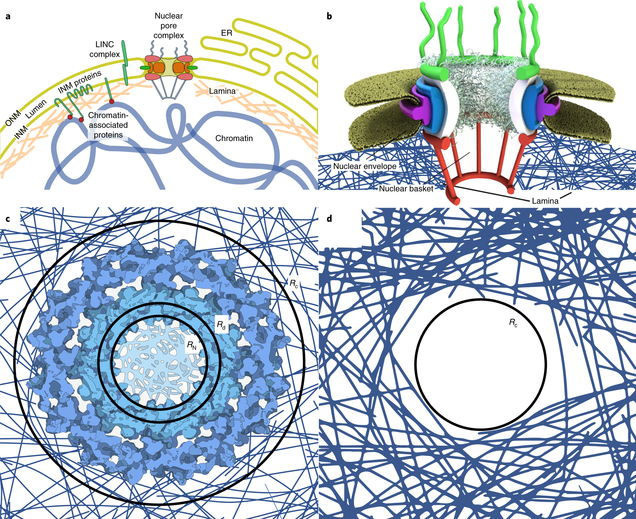
The nuclear lamina consists of two components lamins and nuclear lamin associated membrane proteins. The cells were fixed in 100 methanol for 6 minutes at 20. The quantification of lamin b bands b reveals a significantly higher level of lamin b proteins in total t and nuclear n protein fractions of u118mg and gli1 cells compared to nha cells c. Wild type hela cell lysate lane 4.
2000 j struct biol 129 313 23. The nuclear lamina consists of a two dimensional matrix of proteins located next to the inner nuclear membrane.

